Among other things, Kerri and I were responsible for being “room captains” of the Adults with Type 1 room (sponsored by Insulet (thank you!)) at this year’s Friends for Life conference in Orlando, FL last week.
Being a room captain means introducing speakers, making sure there is water and glucose tabs available, answering questions, and basically making sure the presenters have everything they need before presenting.
I tried hard to fulfill my duties most of the day but was promptly kicked out of the “Pregnancy and Momhood with Type 1” session. Something about upsetting the vibe or some such nonsense.
 Earlier in the day, Jeff Hitchcock explained that Dr. Ben Szirth and his team from the Institute of Ophthalmology and Visual Science at the New Jersey Medical School were doing retinal screenings again, and had brand new equipment that could get extremely detailed images of the eye without the need for dilation (a normal part of a typical diabetes eye exam that turns the exam into a half-day ordeal).
I was very interested, but they were super busy. None of the time slots left in their appointment book worked with my conference schedule. If I had any chance of getting in, it would be during this little pocket of “down time” in my day, and only if they had time for a walk-in. I was blessed and they were able to squeeze me in.
There were a number of stations in the room, each serving a different purpose. My first stop was with an ophthalmology student named Iris. No, I’m not kidding. Iris was checking my irises! I thought that alone deserved a blog post! If she keeps up on her studies, she’s going to be a hit in this field.
She measured my pupils, checked my depth perception (which was a surprisingly difficult test), then checked my vision using the standard “read the smallest line you can see clearly” chart.
Up next was some checks of my vitals (blood pressure, oxygen levels) and another vision test – but this time with a fancy machine. All I had to do was look at a picture of a barn in a field on a screen inside the machine, for a few seconds on each eye, and that was it!
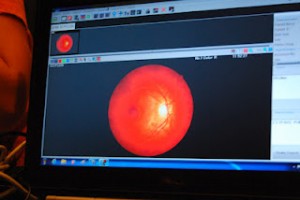
The next station took some seriously fancy scans of the thickness of the back surface of my eyes. It took only seconds on each eye for the computer to grab twenty-some images and do a bunch of fancy measurements and such. If you haven’t noticed already, I have no idea what this machine really looked for, but it was cool nonetheless. Kelly and Brianna have some great pictures of what the docs see on the computer screen – head over to her blog to have a better look.

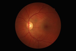
The last station was with Dr. Ben himself. The expert of experts, leading this incredible group of medical students through some advanced diabetes eye health, real-world experience. At this station, Dr. Ben takes a few super-high-res photographs of the back surface of the eye. This allows a very in-depth analysis of the retina and allows him to check for any signs of diabetes-related issues.
He gave me a glowing report, with not even a trace of diabetes-related issues on the retina. In fact, one of the things he said will stick with me for the year…
“I can see from your eyes that you exercise a lot!”
Wow! I have never felt more proud of myself for all of the hard work I put in with my diabetes management, at the gym, and on the bike. Ok, the gym part feels a lot more like fun (basketball!), but still.
The only issue that Dr. Ben saw was some signs of cataract in one eye. He said that is pretty normal for most people, but appear slightly faster for those living with diabetes. Sunglasses are the best form of protection, and can slow the progress quite a bit. He said it might be another twenty years before anything needs to be done about it, and if I’m good about wearing my sunglasses I might stretch that out another ten years.
The images I saw of my eyes and their vascular system were clean, thick, and strong. And I’ve never received a pat on the back that felt better than the words from Dr. Ben…
“I can see from your eyes that you exercise a lot!”


